|
Concept for Proposed Webpage
The first two links to Science (just below) for 11 October, along with others given further down the page, suggest to me the possibility of a story to be told, in laymen's language if possible.
The following points should be addressed:
1. Scientists build upon the work of earlier studies. Several research papers mentioned here span five years, each suggesting possibilites for future work that has been done.
2. A series of fossils may show us the results of evolution (changes in jaw structure, or the adaptation of bones for new purposes) without being able to explain how
such changes were made possible.
3. The popular conception is that evolutionary change must occur through many insensibly fine steps and not by rapid modification of one or a few genes.
4. Modern genetic research permits the kind of experiments that can tell us not only how something might have happened hundreds of millions of years ago, but
also how rapidly major events might have ocurred.
5. Each advance in research leads to predictions that can be confirmed by additional studies.
6. Additional studies often confirm past work and continue the process of discovery and prediction, further confirming evolutionary theory and solidifying our base of knowledge.
I sense that the papers cited below provide the material to tell this story, but I do not have the necessary background to (1) properly evaluate the situation, or (2) to convert the
technical jargon into language that would be convincing to a non-technical reader.
I invite anyone who has the competancy that I lack, and who also has an interest in advancing the comprehension by laymen of evolution, to consider undertaking the explanation of this material.
Illustrations shown below, or those from other sources may be used. I will obtain permissions where necessary.
|
|
Hox and Other Genes Control Large Development Patterns
|
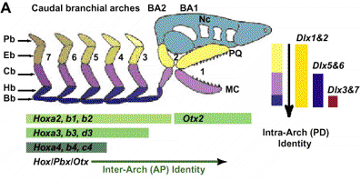
Hypothesized role of Dlx genes in BA patterning. (A) Diagram of a proto-gnathostome neurocranium (Nc) and associated BA (1 to 7) skeletal derivatives.
Gnathostome BA are metameric structures within which develop a proximodistal series of skeletal elements. Inter-BA identity is regulated by Hox, Pbx, and Otx genes.
It is hypothesized that the nested expression of Dlx genes regulates intra-BA identity. (B) In situ hybridization of Dlx2 and Dlx5 (E10.5) and diagram highlighting the
nested Dlx expression within BA mesenchyme. AP, anteroposterior; BA, branchial arch; BA1, first branchial arch; BA2, second branchial arch; Bb, basibranchial; Cb, ceratobranchial;
Eb, epibranchial; Hb, hypobranchial; hy, hyoid arch; md, mdBA1; mx, mxBA1; ; Pb, pharyngeobranchial; PD, proximodistal. From Michael J. Depew, M. J. et al.,
Specification of Jaw Subdivisions by Dlx Genes. Science, 298 (5592) :381-385, October 11, 2002
|
|
Suppression of Control Genes Produces Ancestral Structures
|
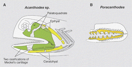
A more symmetrical smile. (A) Skeleton of the head of a jawed vertebrate, Acanthodes, showing symmetry between upper and lower jaw elements (green) and hyoid elements (yellow).
The upper jaw (palatoquadrate) and lower jaw are both subdivided in two by a cartilaginous bridge. (B) Jaws of the acanthodian Poracanthodes with symmetrical jaws and dentition (yellow).
The success of jawed vertebrates is partly attributable to the morphological independence (that is, asymmetry) of upper and lower jaws encoded by differentially expressed Dlx genes.
Elaboration of this developmental code imbued vertebrates with hearing machinery and a wide variety of feeding capabilities. From Koentges G. and T. Matsuoka, Enhanced:
Jaws of the Fates. Science 298 (5592) :371-373, 11 Oct 2002. (requires subscription to see full article).
The Following two paragraphs are also from this Perspectives article.
Jawed vertebrates have three pairs of Dlx homeobox genes--Dlx1/2, Dlx5/6, and Dlx3/7--that are expressed in restricted domains across the proximodistal axis of the branchial arches.
Their nested expression within the branchial arches and the fact that their Drosophila homolog distalless [HN11] is a master regulator of distal leg identity make the
Dlx genes excellent candidates for encoding distal identity in vertebrates. In all bilateral organisms, distalless genes appear to be involved in controlling the outgrowth of body appendages.
Thus, the idea that the vertebrate Dlx homologs serve a similar function is attractive. Rather disappointingly, mice missing a single Dlx gene exhibit only piecemeal changes in the
identities of isolated skeletal elements and teeth. This finding suggested that Dlx genes act as "micromanagers" rather than as "master regulators." Now, Depew and colleagues report the
striking phenotype of the Dlx5/6 double mutant mouse [HN12]. They provide evidence that Dlx5 and Dlx6 are indeed the selectors of distal branchial arch identity.
Their work suggests that the absence of a clear phenotype in mice lacking one Dlx gene is due to compensation by other coexpressed Dlx genes. Thanks to their discovery, the concept of a
proximodistal molecular identity code is alive and well.
The Depew et al. work suggests that lower jaw patterning that is dependent on Dlx5/6 expression may have been elaborated and embellished between the phylogenetic nodes of jawed vertebrate
and bony fish ancestors. Going back one step further in evolutionary history, jawless vertebrates such as lampreys [HN18] only have four Dlx genes with unclear homologies
to their jawed vertebrate counterparts. All lamprey Dlx genes are expressed in branchial arches, but a nested expression pattern appears to be the invention of the jawed vertebrates.
Our knowledge of the enhancer organization that controls this nested Dlx gene expression in jawed vertebrates is still rudimentary. Comparative genomic and functional studies of the
regulatory elements controlling Dlx gene expression in lampreys, sharks, bony fish, coelacanths, and tetrapods will reveal the molecular evolution of the proximodistal code that underlies
the shapes and fates of jaws.
|
|
Control Genes and Early Vertebrate Evolution
|
|
Abstract from Neidert A. et al., Lamprey Dlx Genes and Early Vertebrate Evolution. Proceedings National Academy of Sciences
98 (4) pp. 1665-1670, February 13, 2001.
Gnathostome vertebrates have multiple members of the Dlx family of transcription factors that are expressed during the development of several tissues considered to be vertebrate
synapomorphies, including the forebrain, cranial neural crest, placodes, and pharyngeal arches. The Dlx gene family thus presents an ideal system in which to examine the relationship
between gene duplication and morphological innovation during vertebrate evolution. Toward this end, we have cloned Dlx genes from the lamprey Petromyzon marinus, an agnathan vertebrate
that occupies a critical phylogenetic position between cephalochordates and gnathostomes. We have identified four Dlx genes in P. marinus, whose orthology with gnathostome Dlx genes
provides a model for how this gene family evolved in the vertebrate lineage. Differential expression of these lamprey Dlx genes in the forebrain, cranial neural crest, pharyngeal arches,
and sensory placodes of lamprey embryos provides insight into the developmental evolution of these structures as well as a model of regulatory evolution after Dlx gene duplication events.
|
|
When Important Characters Evolved
|
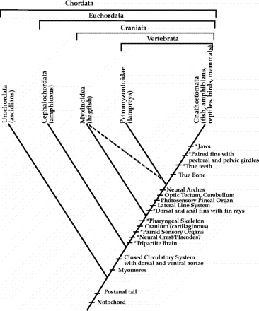
Dlx genes are associated with morphological novelty in the vertebrate lineage. This cladogram depicts hypothesized phylogenetic relationships of extant lineages within the chordates.
A partial listing of morphological characters supporting this phylogenetic hypothesis is shown. Asterisked characters are those that have been shown, in gnathostomes, to be associated
with Dlx gene expression. Note that some analyses using molecular characteristics indicate that hagfish and lampreys are monophyletic (dashed line), suggesting that modern hagfish
secondarily lost certain morphological characters. The position of neural crest and placodes is speculative because hagfish embryos have not been characterized. From Neidert A. et al.,
Lamprey Dlx Genes and Early Vertebrate Evolution. Proceedings National Academy of Sciences 98 (4) :1665-1670, Feb. 13, 2001.
|
|
Vertebrate Jaw Evolution
|

Jaw evolution and functions of signaling molecules in the mandibular arch. (A) Hypothetical evolutionary transition of the jaw. The first two pharyngeal arches [mandibular (ma) and
hyoid (hy) arches] are labeled. Although the arches resemble each other in the ancestral condition, the mandibular arch is enlarged toward gnathostomes (right). Finally, palatoquadrate (pq)
and Meckel's (Mc) cartilages differentiate in the upper and lower jaw, respectively. Only a portion of the published graphic is shown here. From Shigetani Y. et al., Heterotopic
Shift of Epithelial-Mesenchymal Interactions in Vertebrate Jaw Evolution. See Science 296 (5571) 17 May 2002, pp. 1316-1319.
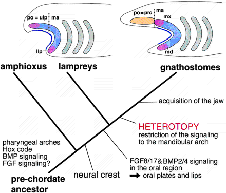
A scenario for jaw evolution. (Top) Patterning of crest cells is compared between lampreys and gnathostomes. In both the animals, the ectomesenchyme contains postoptic (po) and mandibular arch (ma)
subdomains. Growth factors secreted by the epidermis (blue line) induce target homeobox genes (pink and blue) in the entire lamprey ectomesenchyme, whereas in gnathostomes the same signaling is
effective only in the mandibular component. Thus, the phenotypically similar protrusions originate from nonequivalent cell populations in the two animals. Heterotopic shift of epithelial-mesenchymal
interactions is assumed to be behind this difference. The postoptic crest cells occupy the site of prechordal cranium (prc) in gnathostomes. Broken lines indicate the boundary between postoptic (po)
and mandibular (ma) ectomesenchyme. (Bottom) The evolutionary sequence of the changes in developmental patterning is summarized on the phylogenetic tree. The vertebrate ancestor had acquired neural
crest-derived ectomesenchyme after the split from amphioxus, as an element for epithelial-mesenchymal interactions. The molecular cascades for the interactions may have already been present
in the ancestral vertebrates. Although these genes may have already been involved in oral patterning in agnathans, a heterotopic shift must be assumed in the lineage leading to gnathostomes
to explain the topographic discrepancy shown at top. From Shigetani Y. et al., Heterotopic Shift of Epithelial-Mesenchymal Interactions in Vertebrate Jaw Evolution.
See Science 296 (5571) 17 May 2002, pp. 1316-1319.
|
|
The Origin and Evolution of Animal Appendages
|
Another paper that might be usefule in this analysis is: Abstract from Panganiban G. et al., The Origin and Evolution of Animal Appendages.
Proc. Natl. Acad. Sci. USA 94, pp. 5162-5166, May 1997.
Animals have evolved diverse appendages adapted for locomotion, feeding and other functions. The genetics underlying appendage formation are best understood in insects and
vertebrates. The expression of the Distal-less (Dll) homeoprotein during arthropod limb outgrowth and of Dll orthologs (Dlx) in fish fin and tetrapod limb buds led us to examine
whether expression of this regulatory gene may be a general feature of appendage formation in protostomes and deuterostomes. We find that Dll is expressed along the proximodistal
axis of developing polychaete annelid parapodia, onychophoran lobopodia, ascidian ampullae, and even echinoderm tube feet. Dll/Dlx expression in such diverse appendages in these
six coelomate phyla could be convergent, but this would have required the independent co-option of Dll/Dlx several times in evolution. It appears more likely that ectodermal
Dll/Dlx expression along proximodistal axes originated once in a common ancestor and has been used subsequently to pattern body wall outgrowths in a variety of organisms.
We suggest that this pre-Cambrian ancestor of most protostomes and the deuterostomes possessed elements of the genetic machinery for and may have even borne appendages.
|
|
References From Science
|
- Dictionaries and Glossaries
The
On-line Medical Dictionary is available on
CancerWEB.
The
Life Sciences Dictionary is provided by the
BioTech Web site of the University of Texas Institute for Cellular and Molecular Biology.
The
xrefer Web site provides scientific dictionaries and other reference works.
A
Database of Embryological Terms is provided by
M. Cavey, Department of Biological Sciences, University of Calgary, for an
embryology course.
- Web Collections, References, and Resource Lists
Biology Links are provided by the
Dept. of Molecular and Cellular Biology, Harvard University.
The
Google Web Directory provides links to Internet resources related to
developmental biology.
The
Virtual Library-Developmental Biology is provided by the
Society for Developmental Biology.
The
WWW Virtual Library of Cell Biology includes sections of Internet links for
gene expression and
cellular aspects of development.
W. Wasserman, Department of Biology, Loyola University of Chicago, provides a resource page on
developmental biology.
Developmental Biology Online is a supplemental education resource provided by
S. Scadding, Department of Zoology, University of Guelph, Canada. Included are links to Internet resources on
developmental biology and
cell biology and a
glossary. Definitions of
terms for direction and orientation are provided.
- Online Texts and Lecture Notes
J. Kimball provides
Kimball's Biology Pages, an online biology textbook. Three articles on
embryonic development are included.
Embryo Images is a tutorial on mammalian development using scanning electron micrographs provided by the
School of Medicine, University of North Carolina.
The
UNSW Embryology Web site is an educational resource on embryological development provided by
M. Hill, School of Anatomy, University of New South Wales, Australia.
Virtual Embryo/Dynamic Development is a Web resource provided by
L. Browder, Department of Biochemistry and Molecular Biology, University of Calgary.
A
Web supplement to the sixth edition of the textbook Developmental Biology is provided by the author
S. Gilbert, Department of Biology, Swarthmore College, PA. Additional supplemental material is provided on Gilbert's
Zygote Web site.
J. Armbruster, Department of Biological Sciences, Auburn University , provides
lecture notes for a
course on vertebrate comparative anatomy.
L. Zwiebel and
L. Solnica-Krezel, Department of Biological Sciences, Vanderbilt University, provide
lecture notes for a
developmental biology course.
L. Sweeney, Department of Biology, Bryn Mawr College, PA, offers
lecture notes for a
developmental biology course.
G. Podgorski, Department of Biology, Utah State University, offers
lecture notes for a
course on developmental biology.
D. Linden, Biology Department, Occidental College, Los Angeles, offers
lecture notes for a course in
developmental cell biology.
W. Powell, Biology Department, Kenyon College, OH, provides
lecture notes for a
course on genetics and development.
C. Kimmel, Department of Biology, University of Oregon, offers
lecture notes for a
course on vertebrate evolution and development.
D. Rand, Program in Ecology and Evolutionary Biology, Brown University, offers
lecture notes for a
course on evolutionary biology.
- General Reports and Articles
Developmental Dynamics, the journal of the
American Association of Anatomists, makes available an online
collection of review articles.
The
Atlas of Genetics and Cytogenetics in Oncology and Haematology offers a
presentation by J. Bonaventure titled "Skeletal development in human: A model for the study of developmental genes."
The 4 July 1997 issue of Science had a
news article by E. Pennisi and W. Roush titled "Developing a new view of evolution." The 25 June 1999 issue had a
review by A. H. Knoll and S. B. Carroll titled "Early animal evolution: Emerging views from comparative biology and geology."
The May 1997 issue of the
Proceedings of the National Academy of Sciences had an article by
G. Panganiban et al. titled "The origin and evolution of animal appendages." The
25 April 2000 issue had a special feature on evolutionary developmental biology with
perspective and review articles as well as topical
research articles.
Numbered Hypernotes
-
Herman Melville's poem "The Maldive Shark" is available in the
Poetry Archive. D. Campbell, Department of English, Gonzaga University, provides a
resource page about Melville.
-
Sharks and pilot fish.
A photograph by E. Robinson of a
shark with pilot fish is provided on the
ScubaDuba Web site.
An entry for
pilot fish is included in xrefer's
Oxford Paperback Encyclopedia. FishBase has an entry for
Naucrates ductor (pilot fish).
The
NOAA Photo Library displays a 19th-century
etching of a pilot fish.
EnchantedLearning.com offers an educational presentation about
sharks.
The
Ichthyology Department of the Florida Museum of Natural History offers a resource page on
sharks.
Fiona's Shark Mania Web site provides links to
Internet resources on sharks.
-
M. J. Depew and
J. L. R. Rubenstein are in the
Neuroscience Graduate Program and the
Department of Psychiatry, University of California, San Francisco; the
Rubenstein Lab has a Web page.
T Lufkin is at the
Brookdale Center for Developmental and Molecular Biology, Mount Sinai School of Medicine, New York.
-
Homeobox genes Dlx5 and Dlx6.
Homeobox is defined in xrefer's
Dictionary of Biology.
SWISS-PROT has a
keyword entry for
homeobox .
InterPro has an entry for the
homeobox domain.
S. Gilbert makes available a student presentation by K. Panfilio on the
Dlx gene family prepared for a
seminar on developmental genetics.
C. Kimmel offers
lecture notes on Dlx genes for a
course on vertebrate evolution and development.
The
GeneCards database from the
Weizmann Institute of Science has entries for
Dlx5 and
Dlx6 genes with links to other Internet resources provided.
The Jackson Laboratory's
Mouse Genome Informatics Web site has entries for the
Dlx5 and
Dlx6 genes of mice.
-
Congenital disorders possibly related to Dlx genes.
Online Mendelian Inheritance in Man, a catalog of human genes and genetic disorders, has entries for members of the Dlx gene family:
Dlx1, Dlx2,
Dlx3,
Dlx7 (Dlx4),
Dlx5, and
Dlx6.
-
Formation of the vertebrate face.
Embryo Images has a section on
craniofacial development.
T. Marino, Department of Anatomy and Cell Biology, Temple University School of Medicine, provides a presentation on the
development of the face as part of an
embryology resource page.
A student presentation on
human facial development is made available on the
anatomy resource page provided by the
School of Biological Sciences, University of Manchester, UK.
R. Tucker, University of California, Davis, Department of Cell Biology and Human Anatomy, provides lecture
notes on the development of the head, neck, and face (parts
one and two) for a
course on developmental, gross, and radiologic anatomy.
-
Neural crest.
M. Hill's
UNSW Embryology offers a presentation on the
neural crest.
S. Gilbert makes available a student presentation by K. Panfilio on
neural crest cells, which was prepared for a
seminar on developmental genetics.
D. O'Day, Department of Zoology, University of Toronto at Mississauga, provides
lecture notes on the neural crest for a
course on human development.
A student project on the
neural crest was prepared by K. Moran for a
course on developmental biology offered by the School of Biological Sciences, University of Manchester.
The October 1996 issue of Development
had an article
(full text available in PDF format) by G. Koentges and A. Lumsden titled "Rhombencephalic neural crest
segmentation is preserved throughout craniofacial ontogeny" (3).
-
Branchial arches.
Pharyngeal arch (branchial or visceral arch) is defined in xrefer's
Concise Medical Dictionary.
The 1918 edition of
Gray's Anatomy of the Human Body, made available by
Bartleby.com, has a section on the
branchial region.
The
Gross Anatomy Homepage at Marshall University's School of Medicine makes available notes on the
development of branchial arches for a
course on medical gross anatomy.
The
Department of Anatomy, Howard University College of Medicine, makes available lecture notes on the
branchial apparatus for an
anatomical sciences course.
T. Marino provides a presentation on
pharyngeal (branchial arches) as part of an
embryology resource page.
P. Hunt, Department of Biological Sciences, University of Durham, UK, offers a
research presentation on branchial arches.
-
Proximodistal and other axes of development.
Distal and proximal are defined in the
introduction to anatomy terminology provided by the
Faculty of Medicine, University of Calgary School of Medicine.
L. Solnica-Krezel defines proximodistal and other axes of developing limbs in
lecture notes on limb development for a
course on developmental biology.
UNSW Embryology offers a
presentation on the axes of limb molecular development with a
section on proximal/distal patterning.
-
Hox homeobox genes.
Kimball's Biology Pages includes a
section on the Hox cluster of homeobox genes in the
presentation titled "Embryonic development: Putting on the finishing touches."
UNSW Embryology offers a presentation on
Hox homeobox genes in the section on
molecular development and a
presentation on the development of the rostrocaudal axis.
L. Browder's
Dynamic Development makes available a
presentation by D. Rancourt titled "Establishment of spatial patterns of gene expression during early
vertebrate development: Hox genes."
C. Kimmel provides lecture notes on
Hox genes and anterior-posterior patterning for a
course on vertebrate evolution and development.
D. O'Day offers
lecture notes on limb development and Hox genes for a
course on human development.
W. Powell provides
lecture notes on Hox genes and molecular evolution for a
course on genetics and development.
-
Drosophila homolog distalless.
S. Gilbert's
Zygote Web site includes a presentation on
distalless.
FlyBase offers a report on the
gene Dll (distal-less).
The
Interactive Fly offers a presentation on
Dll, including a section on
evolutionary homologs.
-
Dlx5/6 double mutant mouse.
The Jackson Laboratory's TBASE (Transgenic/Targeted Mutation Database) has an entry for the
Dlx5/6 (-/-) mouse, as well as an
April 2002 Knockout of the Month feature on this mouse with links to Internet resources.
-
Mammalian middle ear.
An
Anatomical Tour of the Ear includes a
description of bones of the human middle ear.
The
vertebrates section of the Palaeos Web site provides an
overview of the ear and a presentation on the
incus.
Promenade 'round the Cochlea, a tutorial on the human ear and auditory system, offers a presentation on the
middle ear.
Embryo Images has a Section on
ear development.
R. Tucker provides lecture notes on the
development of the ear for a
course on developmental, gross, and radiologic anatomy.
-
Evolution of the mammalian jaw and ear.
Animal Diversity Web, a presentation of the University of Michigan Museum of Zoology, offers an article on
mammalian jaws and ears.
S. Carr, Department of Biology, Memorial University of Newfoundland, presents a graphic introduction to the
evolution of the mammalian jaw in
lecture notes on systematics for a
course on the principles of evolution and systematics.
Evolution of the mammalian jaw and ear is discussed in an article by
M. Benton titled "Evidence of evolutionary transitions," which is available on the
Actionbioscience.org Web site.
The 23 April 1999 issue of Science had a
News Focus article by E. Pennisi titled "From embryos and fossils, new clues to vertebrate evolution" that discussed the origin of jaws.
C. Kimmel offers
lecture notes titled "Gills make jaws make ears" for a
course on vertebrate evolution and development.
-
Couly et al. paper. The February 2002 issue of Development had an
article by G. Couly, S. Creuzet, S. Bennaceur, C. Vincent, and N. M. Le Douarin
(at the Institut d'Embryologie Cellulaire et Moléculaire du CNRS et du Collège de France) titled "Interactions between Hox-negative cephalic neural crest cells and the
foregut endoderm in patterning the facial skeleton in the vertebrate head."
-
Endoderm is defined in the
BioTech Life Sciences Dictionary and in xrefer's
Dictionary of Biology.
Developmental Biology Online provides a presentation on the
derivatives of endoderm.
D. Linden offers
lecture notes on the endoderm for a course in
developmental cell biology.
-
Acanthodians. An entry for
Acanthodii (acanthodians) is included in xrefer's
Dictionary of Earth Sciences.
The
Palaeos Web site provides an introduction to
Acanthodii.
J. Armbruster includes a section on
Acanthodii in
lecture notes on fish for a
course on vertebrate comparative anatomy.
-
Lampreys.
The online
Columbia Encyclopedia has an entry for
lamprey.
The
University of California Museum of Paleontology offers an introduction to
lampreys.
The
Tree of Life Web Project offers a presentation on
lampreys.
An
information page on lampreys is provided by the SAREP
Web site at the Department of Natural Resources, Cornell University.
J. Armbruster includes a section on jawless fishes (including
lampreys) in lecture notes for a
course on vertebrate comparative anatomy.
The 17 May 2002 issue of Science had a report
by Y. Shigetani et al. titled "Heterotopic shift of epithelial-mesenchymal interactions in vertebrate jaw evolution" (10).
The 13 February 2001 issue of the
Proceedings of the National Academy of Sciences had an
article by A. H. Neidert, V. Virupannavar, G. W. Hooker, and
J. A. Langeland titled "Lamprey Dlx genes and early vertebrate evolution" (9).
|
